The Driving Force Behind Chromatid Movement In Mitosis
The mechanism that moves the chromatids during mitosis is known as the mitotic spindle.
The mitotic spindle is a complex structure composed of microtubules, which are long, thin protein filaments. During mitosis, the mitotic spindle forms between the two poles of the cell and attaches to the chromosomes at their centromeres. The spindle fibers then shorten, pulling the chromosomes to opposite poles of the cell. This process ensures that each daughter cell receives a complete set of chromosomes.
The mitotic spindle is essential for accurate chromosome segregation during mitosis. If the spindle is not formed properly or if the spindle fibers do not function correctly, the chromosomes may not be separated properly, which can lead to aneuploidy, a condition in which cells have an abnormal number of chromosomes. Aneuploidy can have a variety of negative consequences, including developmental disorders, intellectual disability, and cancer.
The mitotic spindle is a highly dynamic structure that undergoes a number of changes during mitosis. These changes are regulated by a variety of proteins, including motor proteins, which move along the microtubules, and regulatory proteins, which control the activity of the motor proteins. The precise coordination of these proteins is essential for the proper functioning of the mitotic spindle and for accurate chromosome segregation.
What Moves the Chromatids During Mitosis
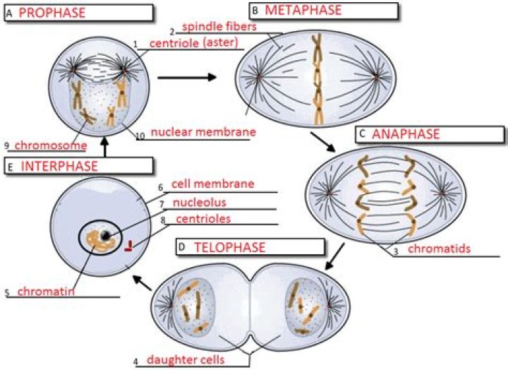
Mitosis is the process by which cells divide, and it is essential for growth, development, and repair. During mitosis, the chromosomes are duplicated and then separated into two new cells. The structure that moves the chromatids during mitosis is the mitotic spindle.
- Chromosomes: The structures that contain the genetic material.
- Chromatids: The two identical copies of each chromosome.
- Centromere: The region of the chromosome where the spindle fibers attach.
- Kinetochore: The protein complex that assembles at the centromere and serves as the attachment point for the spindle fibers.
- Microtubules: The long, thin protein filaments that make up the spindle fibers.
- Motor proteins: The proteins that move along the microtubules, pulling the chromosomes to opposite poles of the cell.
- Regulatory proteins: The proteins that control the activity of the motor proteins.
- Kinetochore checkpoints: The mechanisms that ensure that the chromosomes are properly attached to the spindle fibers before anaphase begins.
- Anaphase: The stage of mitosis during which the chromosomes are separated.
- Telophase: The stage of mitosis during which the spindle fibers disassemble and the two new cells are formed.
The mitotic spindle is a highly dynamic structure that undergoes a number of changes during mitosis. These changes are regulated by a variety of proteins, including motor proteins, which move along the microtubules, and regulatory proteins, which control the activity of the motor proteins. The precise coordination of these proteins is essential for the proper functioning of the mitotic spindle and for accurate chromosome segregation.
Errors in chromosome segregation can lead to aneuploidy, a condition in which cells have an abnormal number of chromosomes. Aneuploidy can have a variety of negative consequences, including developmental disorders, intellectual disability, and cancer.
Chromosomes
Chromosomes are the structures that contain the genetic material of a cell. They are made up of DNA, which is a long, thin molecule that contains the instructions for making all of the proteins in the cell. During mitosis, the chromosomes are duplicated and then separated into two new cells. The structure that moves the chromatids during mitosis is the mitotic spindle.
The mitotic spindle is a complex structure composed of microtubules, which are long, thin protein filaments. During mitosis, the mitotic spindle forms between the two poles of the cell and attaches to the chromosomes at their centromeres. The spindle fibers then shorten, pulling the chromosomes to opposite poles of the cell. This process ensures that each daughter cell receives a complete set of chromosomes.
The proper segregation of chromosomes during mitosis is essential for the survival of the cell. If the chromosomes are not separated properly, the daughter cells will not have the correct number of chromosomes, which can lead to a variety of problems, including developmental disorders, intellectual disability, and cancer.
The connection between chromosomes and the mitotic spindle is essential for the proper division of cells. The chromosomes provide the genetic material that is essential for the survival of the cell, while the mitotic spindle provides the structure that moves the chromosomes during mitosis. This process ensures that each daughter cell receives a complete set of chromosomes, which is essential for the survival of the organism.
Chromatids
Chromatids are the two identical copies of each chromosome that are created during DNA replication. They are joined together at the centromere, which is a specialized region of the chromosome. During mitosis, the mitotic spindle attaches to the centromeres of the chromosomes and pulls the chromatids apart, moving them to opposite poles of the cell. This process ensures that each daughter cell receives a complete set of chromosomes.
The connection between chromatids and the mitotic spindle is essential for the proper division of cells. If the chromatids are not separated properly, the daughter cells will not have the correct number of chromosomes, which can lead to a variety of problems, including developmental disorders, intellectual disability, and cancer.
The proper segregation of chromatids during mitosis is essential for the survival of the cell. This process ensures that each daughter cell receives a complete set of chromosomes, which is essential for the survival of the organism.
Centromere
The centromere is a specialized region of the chromosome that plays a critical role in chromosome segregation during mitosis. It is the site where the spindle fibers attach to the chromosomes, and it is essential for the proper movement of the chromosomes during cell division.
The centromere is composed of a complex of proteins that form a kinetochore, which is the structure that binds to the spindle fibers. Once the spindle fibers are attached to the kinetochore, they shorten, pulling the chromosomes to opposite poles of the cell. This process ensures that each daughter cell receives a complete set of chromosomes.
The centromere is essential for the proper segregation of chromosomes during mitosis. If the centromere is not properly formed or if the spindle fibers do not attach to the kinetochore, the chromosomes may not be separated properly, which can lead to aneuploidy, a condition in which cells have an abnormal number of chromosomes. Aneuploidy can have a variety of negative consequences, including developmental disorders, intellectual disability, and cancer.
The centromere is a complex and dynamic structure that is essential for accurate chromosome segregation during mitosis. Understanding the structure and function of the centromere is critical for understanding the process of cell division and for developing new treatments for aneuploidy.
Kinetochore
The kinetochore is a protein complex that assembles at the centromere of each chromosome during mitosis. It serves as the attachment point for the spindle fibers, which are responsible for moving the chromosomes to opposite poles of the cell. The kinetochore is essential for the proper segregation of chromosomes during mitosis, and defects in kinetochore function can lead to aneuploidy, a condition in which cells have an abnormal number of chromosomes.
The kinetochore is a complex structure that is composed of over 100 different proteins. These proteins work together to form a platform that binds to the spindle fibers. The kinetochore also contains motor proteins that move along the spindle fibers, pulling the chromosomes to opposite poles of the cell.
The kinetochore is essential for the proper segregation of chromosomes during mitosis. If the kinetochore is not properly formed or if the spindle fibers do not attach to the kinetochore, the chromosomes may not be separated properly, which can lead to aneuploidy. Aneuploidy can have a variety of negative consequences, including developmental disorders, intellectual disability, and cancer.
The kinetochore is a complex and dynamic structure that is essential for accurate chromosome segregation during mitosis. Understanding the structure and function of the kinetochore is critical for understanding the process of cell division and for developing new treatments for aneuploidy.
Microtubules
Microtubules are long, thin protein filaments that make up the spindle fibers, which are responsible for moving the chromatids during mitosis. Microtubules are composed of a protein called tubulin, which polymerizes to form long, hollow cylinders. The spindle fibers are formed by the polymerization of microtubules at the two poles of the cell. The spindle fibers then attach to the kinetochores of the chromosomes, which are protein complexes that assemble at the centromere of each chromosome.
- Structure of microtubules: Microtubules are composed of a protein called tubulin, which polymerizes to form long, hollow cylinders. The tubulin subunits are arranged in a spiral pattern, which gives microtubules their characteristic strength and flexibility.
- Assembly of spindle fibers: Spindle fibers are formed by the polymerization of microtubules at the two poles of the cell. The microtubules polymerize in a dynamic manner, with new subunits constantly being added and removed from the ends of the fibers. This dynamic instability allows the spindle fibers to search for and attach to the kinetochores of the chromosomes.
- Attachment of spindle fibers to kinetochores: The spindle fibers attach to the kinetochores of the chromosomes, which are protein complexes that assemble at the centromere of each chromosome. The kinetochores are composed of a number of different proteins, which work together to bind to the spindle fibers and ensure that the chromosomes are properly aligned and segregated during mitosis.
- Movement of chromosomes: Once the spindle fibers are attached to the chromosomes, they shorten, pulling the chromosomes to opposite poles of the cell. The shortening of the spindle fibers is driven by motor proteins, which move along the microtubules, pulling the chromosomes with them. This process of chromosome movement is essential for the proper segregation of chromosomes during mitosis.
Microtubules are essential for the proper segregation of chromosomes during mitosis. Defects in microtubule function can lead to aneuploidy, a condition in which cells have an abnormal number of chromosomes. Aneuploidy can have a variety of negative consequences, including developmental disorders, intellectual disability, and cancer.
Motor proteins
Motor proteins are essential for the proper segregation of chromosomes during mitosis. They move along the microtubules of the mitotic spindle, pulling the chromosomes to opposite poles of the cell. This process ensures that each daughter cell receives a complete set of chromosomes.
- Kinesin is a motor protein that moves towards the plus end of microtubules. It is responsible for pulling the chromosomes to the poles of the cell during mitosis.
- Dynein is a motor protein that moves towards the minus end of microtubules. It is responsible for pulling the poles of the cell apart during mitosis.
- Cytoplasmic dynein is a motor protein that moves along microtubules in the cytoplasm. It is responsible for pulling organelles and vesicles around the cell.
- Myosin is a motor protein that moves along actin filaments. It is responsible for muscle contraction and cell movement.
Motor proteins are essential for a variety of cellular processes, including mitosis, cell movement, and organelle transport. Defects in motor protein function can lead to a variety of diseases, including cancer and neurodegenerative disorders.
Regulatory proteins
Regulatory proteins are essential for the proper segregation of chromosomes during mitosis. They control the activity of the motor proteins that move the chromosomes along the spindle fibers. Without regulatory proteins, the motor proteins would not be able to function properly and the chromosomes would not be able to move to opposite poles of the cell. This would lead to aneuploidy, a condition in which cells have an abnormal number of chromosomes. Aneuploidy can have a variety of negative consequences, including developmental disorders, intellectual disability, and cancer.
One of the most important regulatory proteins is called Mad2. Mad2 binds to the kinetochore of each chromosome and prevents the motor proteins from attaching to the spindle fibers until all of the chromosomes are properly aligned. Once all of the chromosomes are aligned, Mad2 releases the motor proteins and they begin to move the chromosomes to opposite poles of the cell.
Regulatory proteins are essential for the proper segregation of chromosomes during mitosis. Defects in regulatory protein function can lead to aneuploidy, a condition that can have a variety of negative consequences. Understanding the function of regulatory proteins is critical for understanding the process of mitosis and for developing new treatments for aneuploidy.
Kinetochore checkpoints
Kinetochore checkpoints are essential for the proper segregation of chromosomes during mitosis. They ensure that all of the chromosomes are properly attached to the spindle fibers before anaphase begins. This is important because if even one chromosome is not properly attached, it can lead to aneuploidy, a condition in which cells have an abnormal number of chromosomes. Aneuploidy can have a variety of negative consequences, including developmental disorders, intellectual disability, and cancer.
The kinetochore checkpoint is a complex process that involves a number of different proteins. One of the most important proteins is Mad2. Mad2 binds to the kinetochore of each chromosome and prevents the motor proteins from attaching to the spindle fibers until all of the chromosomes are properly aligned. Once all of the chromosomes are aligned, Mad2 releases the motor proteins and they begin to move the chromosomes to opposite poles of the cell.
Kinetochore checkpoints are essential for accurate chromosome segregation during mitosis. Defects in kinetochore checkpoint function can lead to aneuploidy, a condition that can have a variety of negative consequences. Understanding the function of kinetochore checkpoints is critical for understanding the process of mitosis and for developing new treatments for aneuploidy.
Anaphase
Anaphase is a critical stage of mitosis, the process by which cells divide. During anaphase, the chromosomes, which carry the cell's genetic information, are separated and pulled to opposite poles of the cell. This process is essential for ensuring that each daughter cell receives a complete set of chromosomes.
What moves the chromatids during mitosis? The movement of the chromosomes during anaphase is driven by a complex interplay of proteins and structures within the cell.
- Kinetochore microtubules: These microtubules attach to the kinetochore, a protein complex located at the centromere of each chromosome. The kinetochore microtubules are responsible for pulling the chromosomes to opposite poles of the cell.
- Motor proteins: Motor proteins are proteins that move along microtubules. During anaphase, motor proteins use the energy from ATP to move towards the poles of the cell, pulling the kinetochore microtubules and the attached chromosomes with them.
- Regulatory proteins: Regulatory proteins control the activity of motor proteins. During anaphase, regulatory proteins ensure that the motor proteins move in the correct direction and at the correct speed.
The precise coordination of these proteins and structures is essential for the proper segregation of chromosomes during anaphase. Errors in chromosome segregation can lead to aneuploidy, a condition in which cells have an abnormal number of chromosomes. Aneuploidy can have a variety of negative consequences, including developmental disorders, intellectual disability, and cancer.
Telophase
Telophase is the final stage of mitosis, the process by which cells divide. During telophase, the spindle fibers disassemble and the two new cells are formed. This process is essential for ensuring that each daughter cell receives a complete set of chromosomes.
The movement of the chromosomes during mitosis is driven by a complex interplay of proteins and structures within the cell. The spindle fibers, which are composed of microtubules, play a critical role in this process. During anaphase, the spindle fibers shorten, pulling the chromosomes to opposite poles of the cell. Once the chromosomes reach the poles, the spindle fibers disassemble and the two new cells are formed.
Telophase is a critical stage of mitosis, as it ensures that each daughter cell receives a complete set of chromosomes. Errors in chromosome segregation can lead to aneuploidy, a condition in which cells have an abnormal number of chromosomes. Aneuploidy can have a variety of negative consequences, including developmental disorders, intellectual disability, and cancer.
The connection between telophase and the movement of the chromatids during mitosis is essential for understanding the process of cell division. By understanding this connection, scientists can gain a better understanding of how cells divide and how errors in cell division can lead to disease.
FAQs
What is mitosis?
Mitosis is the process by which cells divide. It is essential for growth, development, and repair.
What are chromatids?
Chromatids are the two identical copies of each chromosome that are created during DNA replication.
What moves the chromatids during mitosis?
The chromatids are moved during mitosis by the mitotic spindle, a complex structure composed of microtubules.
What is the role of the mitotic spindle?
The mitotic spindle forms between the two poles of the cell and attaches to the chromosomes at their centromeres. The spindle fibers then shorten, pulling the chromosomes to opposite poles of the cell.
What happens if the mitotic spindle does not function properly?
If the mitotic spindle does not function properly, the chromosomes may not be separated properly, which can lead to aneuploidy, a condition in which cells have an abnormal number of chromosomes.
What are the consequences of aneuploidy?
Aneuploidy can have a variety of negative consequences, including developmental disorders, intellectual disability, and cancer.
Summary:
The movement of the chromatids during mitosis is essential for the proper segregation of chromosomes. The mitotic spindle is a complex structure that plays a critical role in this process.
Next Article Section:
The Importance of Mitosis
Tips for Understanding "What Moves the Chromatids During Mitosis"
Understanding the process of mitosis, particularly the movement of chromatids, is crucial for comprehending cell division and genetics. Here are some tips to enhance your understanding:
Tip 1: Visualize the Process
Visual aids such as diagrams, animations, and videos can greatly enhance your understanding of mitosis. These resources can illustrate the complex interactions of proteins and structures involved in chromatid movement.
Tip 2: Learn the Terminology
Familiarize yourself with the key terms related to mitosis, such as chromatids, centromeres, microtubules, and motor proteins. Understanding these terms will enable you to follow scientific discussions and research papers on the topic.
Tip 3: Study the Mitotic Spindle
The mitotic spindle is the primary structure responsible for moving chromatids. Delve into its composition, dynamics, and the role of its components, including microtubules, motor proteins, and regulatory proteins.
Tip 4: Explore the Molecular Mechanisms
Understand the molecular mechanisms underlying chromatid movement. Research the interactions between motor proteins, regulatory proteins, and microtubules, and how these interactions drive the precise movement of chromatids.
Tip 5: Analyze Scientific Literature
Refer to scientific journals and articles to stay updated on the latest research and advancements in the field. Reading scientific literature will deepen your knowledge and provide insights into the ongoing exploration of mitosis.
Summary:
By following these tips, you can gain a comprehensive understanding of the mechanisms that move chromatids during mitosis. This knowledge is essential for unraveling the complexities of cell division and its implications in biology and medicine.
Next Article Section:
The Significance of Mitosis in Cell Biology
Conclusion
Throughout this article, we have explored the intricate mechanisms that drive the movement of chromatids during mitosis. This process, facilitated by the mitotic spindle, is essential for the accurate segregation of genetic material during cell division.
Understanding the molecular players and their precise interactions has profound implications for cell biology and medicine. Mitosis is a fundamental process that underpins growth, development, and tissue repair. Dysregulation of mitosis can lead to aneuploidy, a condition associated with developmental disorders, infertility, and cancer. Therefore, deciphering the mechanisms that govern chromatid movement is crucial for unraveling the causes and potential treatments for these conditions.
Unveiling The Extraordinary Journey Of Trevor Noah's Mother
Unveiling The Luminescent Genius Of Patricia Azarcoya Schneider
Unveiling The Heights Of Dominic Purcell: A Journey Of Discovery